
BONLAB BLOG
Thoughts
&
Scientific Fiction

Fibers made by assembly of emulsion droplets
Fibers are interesting. They are made by a spinning process in which a liquid based mixture, referred to as spinning dope, is extruded through an orifice hereby generating a jet, which subsequently is solidified through either coagulation/precipitation and/or gelation. Two extreme fibers found in Nature are spidersilk, a super strong and extensible liquid-crystalline fiber, and the soft hydrogel double-strings of toad eggs, as spawn by the common toad (Bufo bufo). The production of manmade fibers using dry and wet spinning techniques – both starting from a liquid mixture – goes back to the 19th century. An early example is the development of Rayon fibers initiated by the discovery of Schweizer in 1857, who found that cellulose could be dissolved in and re-precipitated from an aqueous solution of ammonia and copper (II) hydroxide (coined Schweizer’s reagent (dry or wet)). Examples of wet-spun high performance fibers include ultrahigh molecular weight poly(ethylene) fibers, and polyaramid fibers.
An emerging trend is to make soft, hydrogel-based, fibers wet spun into water. Applications for example are in the area of tissue engineering. Microfluidic technologies are often employed to manufacture these fibers.
We asked ourselves whether it would be possible to fabricate fibers through assembly of thousands of emulsion droplets? We call these HIPE (High Internal Phase Emulsion) fibers.

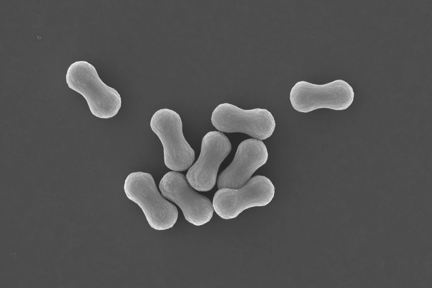
Fig. (a) Schematic representation of the fabrication of microfluidically spun HIPE fiber. The diagram shows coaxial flow channels in the microfluidic device, where the inner and outer capillaries contain concentrated emulsion and acidic water, respectively, in a continuous flow. A continuous HIPE fiber is produced upon exposure of the emulsion to the acidic water. The acidic conditions allow the formation of multiple hydrogen bonds between emulsion droplets, initiating them to assemble into a macroscopic supracolloidal fiber. (b) Photograph of a HIPE fiber in acidic aqueous solution (top left), and disintegration into individual dispersed emulsion droplets upon addition of base (top right). Scale bars represent 1 cm. Light micrographs of the HIPE fiber in acidic condition (bottom left) and disintegrated HIPE fiber in basic condition (bottom right). Scale bars represent 50 µm and 25 µm, respectively. (c) Photograph of HIPE fibers with uniform length. The HIPE fibers with uniform length were fabricated by utilizing air bubbles as a cutting mechanism. Scale bar represents 0.5 cm. (d) Photograph of asymmetric Janus HIPE fiber. Scale bars represent 0.1 cm (top) and 300 µm (bottom). (e) Photograph of asymmetric HIPE fiber consisting of three different sections ‘toothpaste’. Scale bars represent 0.1 cm (top) and 300 µm (bottom). (f) Photograph of magnetic HIPE fiber attracted by an external magnet (fiber with a slight yellow color - left hand side), and no magnetic response with the non-magnetic HIPE fiber (white color fiber - right hand side). Scale bar represents 1 cm.
In our paper published in the Journal of Materials Chemistry A we show the fabrication of fibers from emulsion droplets. We use flow-focussing microfluidic set-up whereby a generated jet of emulsion droplets stabilized by amphiphilic pH-responsive branched copolymers (pH-BCP) and reinforced by Laponite clay discs is exposed to an external surrounding liquid flow of lower pH. Proton diffusion into the stream of emulsion droplets induces the self-assembly process leading to the formation of a continuous supracolloidal fiber. We demonstrate that the fiber can disintegrate back into an oil-in-water emulsion. We discuss control of fiber composition hereby using two and three combined streams of emulsion droplets to generate Janus fibers, and using ferrofluids to produce magnetic fibers. We show control of fiber length by employing air bubbles as a mean to produce short fibers of discrete length. Looking towards applications we demonstrate the use of our supracolloidal emulsion droplet fibers as a material to control the delivery of volatile compounds through evaporation, and we show that the dried fibers are a nanocomposite highly porous and light material.
You can read the paper here: http://dx.doi.org/10.1039/C5TA08917D

Corinna Preuss awarded Newton International Fellowship
Dr Corinna Preuss has been awarded a Newton International Fellowship to conduct research at the University of Warwick’s Department of Chemistry.
Corinna Preuss
Jointly run by The British Academy, The Academy of Medical Sciences and the Royal Society, the Fellowship is for non-UK scientists who are at an early stage of their research career and provides the opportunity for the best early stage post-doctoral researchers from all over the world to work at UK research institutions for a period of two years.
Speaking after being awarded the Fellowship Dr Preuss said:
“I’m very honoured and delighted to be awarded the Newton International Fellowship. Not only will it support my personal development but is also emphasises the novelty and importance of our proposed research project.”
Dr Preuss will work as part of a team led by Professor Stefan Bon to mimic the motional behaviour of zooplankton by fabricating artificial jelly-objects that have the capability to transform shape, swim, and – as an additional feature – release payloads. Dr Preuss says these hydrogel objects will have these three pre-programmed functions “which can be triggered on demand in a controlled fashion”. For this purpose, recent scientific advances in polymer and colloid chemistry will be merged with soft matter physics and robotics in order to create a promising and interdisciplinary research program
Further to the research with Professor Bon, Dr Preuss is keen to use the Fellowship to teach undergraduate chemists and to create a network with other fellow scientists, saying that: “In my opinion, exchanging knowledge and listening to different opinions is essential for the formation of a highly efficient scientific society”.
Discussing why she chose the University of Warwick Dr Preuss said:
“I met Professor Stefan Bon during a conference in Mexico.I was impressed by his research and the passion he presented it with. Later on, whilst I was presenting my research at the poster session, we got the chance to chat more and discovered that our interests in each other’s research would create a promising base for a further collaboration. In working with Stefan and coming to the University of Warwick, I’m taking the chance of changing my field of research to colloidal chemistry and engineering, which provides a new, challenging and fascinating area for me”.
Contacts:
Tom Frew - International Press Officer
Email: a.t.frew@warwick.ac.uk
Tel: +44 (0)247 657 5910
Mob: +44 (0)7785 433 155
Control of vesicle membrane permeability with catalytic particles
The ability to control membrane permeability in vesicles allows for regulated transport of matter across the vesicular wall. Vesicles can be seen as microscopic sacs containing a compartmentalized volume of liquid dispersed in a bulk liquid environment. Compartmentalization of small and finite volumes of liquid and consecutive the emergence of membrane bioenergetics are identified as being of key importance in the evolution of cells, and hence the origin of life. Nature has devised sophisticated strategies to accomplish control of transmembrane transport, including endo- and exocytosis as well as the incorporation of transmembrane proteins into cell membranes. A variety of synthetic approaches have been explored by scientists in order to accomplish such control in manmade systems. Examples include hybrid systems whereby transmembrane proteins were incorporated as part of synthetic vesicles, and the use of responsive macromolecular building blocks to regulate membrane porosity upon an external trigger in polymer vesicles, also referred to as polymersomes.
The ability to control membrane permeability in vesicles allows for regulated transport of matter across the vesicular wall. Vesicles can be seen as microscopic sacs containing a compartmentalized volume of liquid dispersed in a bulk liquid environment. Compartmentalization of small and finite volumes of liquid and consecutive the emergence of membrane bioenergetics are identified as being of key importance in the evolution of cells, and hence the origin of life. Nature has devised sophisticated strategies to accomplish control of transmembrane transport, including endo- and exocytosis as well as the incorporation of transmembrane proteins into cell membranes. A variety of synthetic approaches have been explored by scientists in order to accomplish such control in manmade systems. Examples include hybrid systems whereby transmembrane proteins were incorporated as part of synthetic vesicles, and the use of responsive macromolecular building blocks to regulate membrane porosity upon an external trigger in polymer vesicles, also referred to as polymersomes.

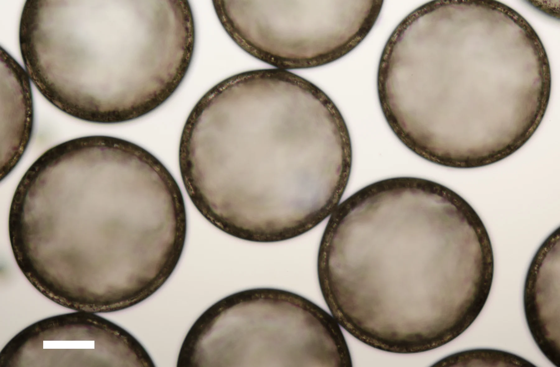
Dark field microscopy image of polymer vesicles which contain manganese oxide colloidal particles in their membranes, pre-loaded with barium ions and dispersed in water (containing sulfate anions) after their exposure to a low external concentration of hydrogen peroxide. The white haze surrounding the vesicles and the white tails are the result of the precipitation of barium sulfate crystals upon release of the barium cations from the vesicles. Scale bar: 100 μm
In our recent paper published in Materials Horizons we show for the first time that the permeability of the membrane of polymer vesicles can be controlled by membrane-embedded catalytically active manganese oxide particles. The ability to chemically trigger activity of the catalytic particle, hereby inducing a temporary increase of membrane permeability, offers precise time-specific control of transmembrane transport. It is our belief that this concept can be applied to a wide variety of membrane-based systems. At the end of the paper we open a disccusion for the origins of life and protocell communities that a hybrid vesicular structure, which has “active” colloidal particles as part of its membrane, may have regulated permeability in primitive cells.
to read the paper: http://dx.doi.org/10.1039/C5MH00093A